Progressive optic neuropathy, usually from elevated intraocular pressure (IOP), leading to characteristic visual field loss and potentially blindness. Two major types exist: open-angle glaucoma (chronic, more common – impaired aqueous drainage despite an open anterior chamber angle) and angle-closure glaucoma (acute – sudden blockage of the angle causing a rapid IOP rise).
Glaucoma is the second leading cause of blindness worldwide. It often goes undetected until late (open-angle is called the "silent thief of sight"), so screening is crucial. Acute angle-closure is an ophthalmologic emergency that can cause permanent vision loss within hours if not promptly treated. Both scenarios are frequently tested, requiring recognition of subtle chronic findings and acute red-flag symptoms.
Open-angle glaucoma: typically painless with insidious peripheral vision loss (patients may not notice gradual "tunnel vision"). Often picked up on routine exam by ↑IOP or optic disc cupping. Risk factors: age >40, African or Hispanic ancestry, family history, diabetes.
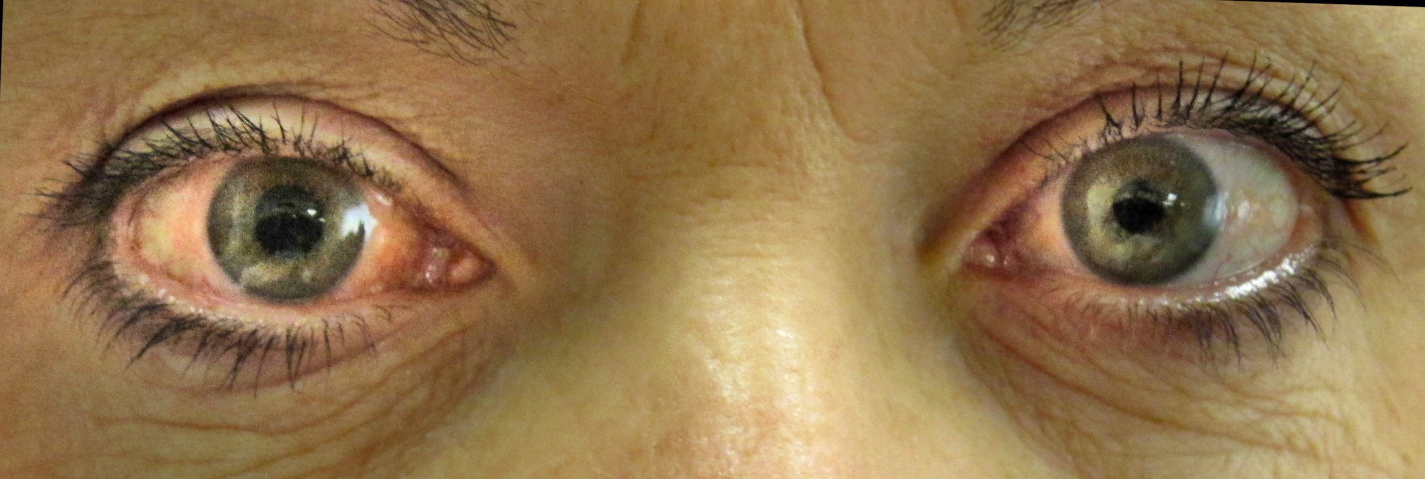
Angle-closure glaucoma: acute onset of severe eye pain with blurred vision and halos around lights; often associated with headache, nausea/vomiting. The affected eye is red with a mid-dilated, fixed pupil and feels hard due to markedly elevated IOP (often 30–50+ mmHg). Commonly triggered by pupillary dilation (e.g. entering a dark room or certain drugs) in a susceptible (usually older, hyperopic) patient.
Measure intraocular pressure (tonometry): Normal ~10–21 mmHg; IOP >21 is a risk factor (though some glaucoma occurs at normal pressure).
Perform fundoscopic exam: look for optic disc cupping (cup-to-disc ratio >0.5 or asymmetry) and thinning of the neural rim, suggesting glaucomatous damage.
Assess visual fields (perimetry): glaucoma causes characteristic peripheral field defects (e.g. arcuate scotomas, nasal step) corresponding to optic nerve damage.
Use gonioscopy (slit-lamp with special lens) to examine the anterior chamber angle – determines if angle is open vs. narrow/closed and helps identify any secondary causes.
If acute angle-closure is suspected, do not wait for detailed testing – initiate immediate IOP-lowering treatment, then perform definitive intervention (laser iridotomy) once the cornea clears.
Condition
Distinguishing Feature
Ocular hypertension
elevated IOP without optic nerve damage or visual field loss (no neuropathy yet)
Acute anterior uveitis
painful red eye with photophobia, but small pupil (miotic), cells in anterior chamber, and usually normal or low IOP
Cataract
painless progressive vision loss due to lens opacity (glare, night vision difficulty), affects central vision first (no cupping or high IOP)
Open-angle (chronic): start with topical drops to lower IOP – typically a prostaglandin analog (e.g. latanoprost) first-line, and/or beta-blocker (timolol); adjuncts include alpha-2 agonists (brimonidine) or carbonic anhydrase inhibitors (dorzolamide). If medical therapy insufficient, use laser trabeculoplasty or surgical trabeculectomy/tube shunt to increase aqueous outflow.
Angle-closure (acute): this is an emergency – immediately give multiple IOP-lowering meds (e.g. topical β-blocker, alpha-agonist, and pilocarpine plus systemic acetazolamide; add osmotic agent like IV mannitol if needed). Definitive treatment is a laser peripheral iridotomy (creating a small hole in the iris) to relieve pupillary block – typically performed in the affected eye once pressure drops, and prophylactically in the fellow eye.
Mnemonic: O-PEN for open-angle glaucoma – Optic disc cupping, Progressive peripheral vision loss, Elevated IOP, No early symptoms.
In acute angle-closure think "painful red eye with halos" – a rock-hard eye with mid-dilated pupil in a patient with sudden vision loss is an ophthalmic emergency.
Signs of acute angle-closure (severe eye pain, blurred vision with halos, vomiting, fixed mid-dilated pupil) → indicates vision-threatening emergency (if IOP isn't lowered within hours, permanent optic nerve damage can occur).
Avoid mydriatic medications in patients with known narrow angles – dilating the pupil can precipitate acute angle-closure glaucoma.
Risk factors or optic disc changes → suspect glaucoma.
Check IOP (tonometry) and optic disc on exam; if abnormal, perform gonioscopy to classify angle and visual field testing to confirm deficits.
Open-angle suspected: manage chronically with drops to control IOP and monitor optic nerve and fields regularly.
Angle-closure suspected: begin immediate IOP-lowering therapy; once pressure improves, perform laser iridotomy to definitively open the angle.
Screen high-risk individuals (older adults, positive family history) with periodic comprehensive eye exams (including IOP measurement and fundoscopy).
Older African American patient with gradual loss of peripheral vision and cupped optic discs on exam (high IOP) → Primary open-angle glaucoma.
Middle-aged hyperopic woman with sudden eye pain after sitting in a dark theater, plus headache, nausea, a red eye and non-reactive mid-dilated pupil → Acute angle-closure glaucoma.
Case 1
A 65‑year‑old African American man reports gradually worsening peripheral vision over the past year.
Case 2
A 55‑year‑old far-sighted woman develops sudden severe right eye pain while watching a movie in a dark theater.
Right eye with acute angle-closure glaucoma (red, injected conjunctiva and mid-dilated pupil).